Latest news from MDF on myotonic dystrophy Type 1 (DM1).
Sign up for newsletter
Learn more
March 18, 2014
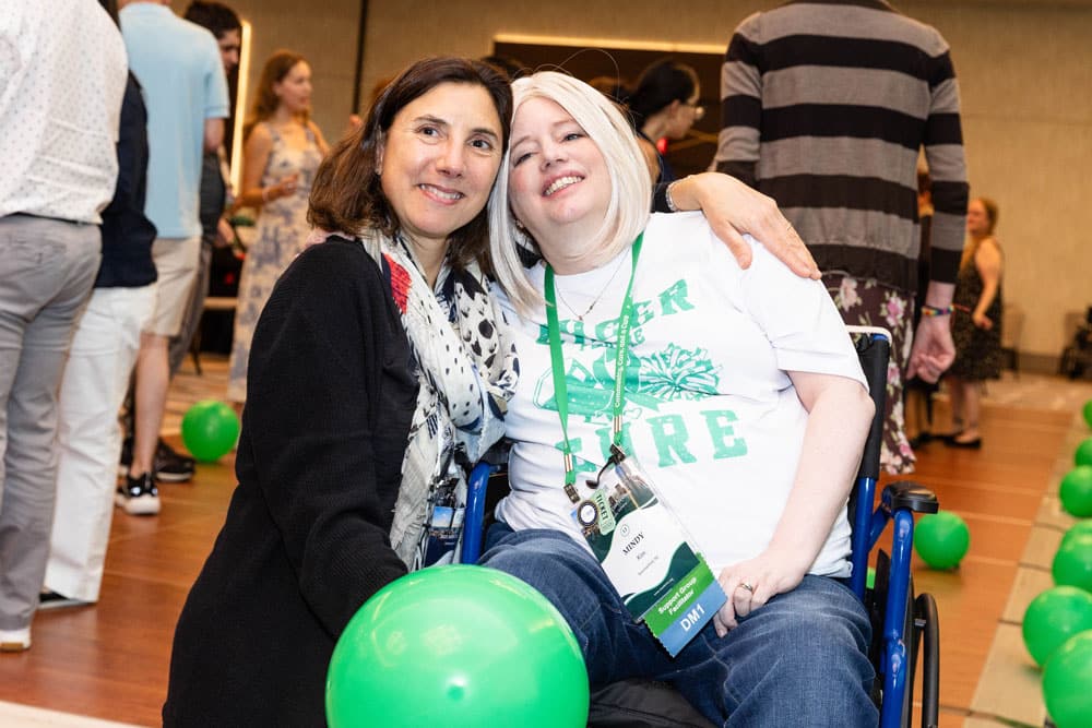
Attend virtual and in-person events that bring the community together.
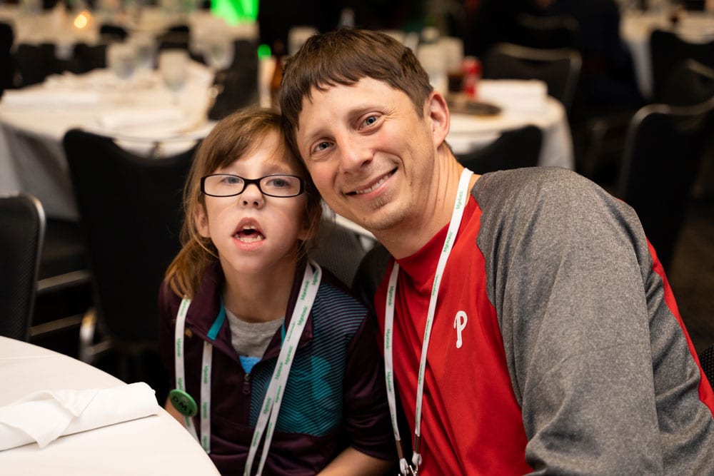
Family stories that reflect the challenges and triumphs of those living with DM.
Make a lasting impact by supporting initiatives for Myotonic Dystrophy.