News  Donate!

Research

Structure of the Myotonic Dystrophy Type 2 RNA

Published on Wed, 01/22/2014

Researchers at important academic labs around the US have recently published exciting new information about advances in DM research. The Matthew Disney Lab at The Scripps Research Institute in Florida announced the results of a study examining RNA toxicity in DM2 patients. Summaries of the studies and results are below, along with links to the PubMed abstracts and complete research publications.

Structure of the Myotonic Dystrophy Type 2 RNA and Designed Small Molecules that Reduce Toxicity Childs-Disney et al (Matthew Disney Lab)

Researchers at Scripps Research Institute in Florida recently examined the structure of the toxic RNA molecule made from the DNA mutation causing myotonic dystrophy type 2 (DM2). Dr. Matthew Disney and his colleagues used a technique called X-ray crystallography to look at the shape of the RNA at the atomic level in order to determine what types of drugs would best attach to it and reduce its toxicity.  

One of the reasons the DM2 RNA is unhealthy in cells is because it changes how other genes are processed and regulated. They showed that their custom-designed drugs were able to reverse the improper processing of a gene known to be affected in DM by varying degrees depending on the design of the drug. This study shows that drugs they previously proposed could bind the toxic RNA are now able to be administered to cells with toxic RNA similar to DM2 and reduce the RNA toxicity.

For more information:

Click here to view a pdf of the full article

Click here to read the abstract

01/22/2014

MDF Grant Recipient Develops Tool for Measuring the Impact of Childhood DM on Quality of Life

Published on Wed, 12/18/2013

To support our commitment to research, MDF awards fellowship grants to postdoctoral researchers as part of our annual Fund-A-Fellow (FAF) program. We recently caught up with one of our current grant recipients, Dr. Nicholas Johnson, Assistant Professor of Neurology at the University of Utah.

Dr. Johnson was inspired to focus his practice and research on myotonic dystrophy after working for five years at the University of Rochester with Dr. Chad Heatwole, Dr. Richard Moxley, Dr. Charles Thornton and a number of other clinicians and researchers leading the charge to find new treatments and cure for DM. He opened his practice at the University of Utah in July of 2013 and is excited to be able to treat the large DM population in Utah and surrounding states.

“There is an excellent neurology department at the University of Utah,” Dr. Johnson says, “but previously no one was focused specifically on researching DM. Being here gives me the opportunity not only to provide ongoing treatment to those individuals, but to include many of them in our research studies as well, which is exciting.”

We spoke with Dr. Johnson about his current FAF research.

MDF: First, tell us about the research you’ve been doing as a result of receiving the FAF grant.

NJ: “Sure. Once I received the FAFA grant, I began a project in March 2012 to develop a tool that can be used in a clinic or in research studies to measure how myotonic dystrophy impacts the quality of life of a child with congenital or childhood-onset DM. 

“The tool is called Congenital and Childhood Myotonic Dystrophy Health Index or CCMDHI. It’s a survey-based tool that collects data about quality of life from birth to age 18. Until now, there’s been very little data about how DM progresses from birth to adulthood, but that information is necessary for clinical trials to move forward.”

MDF: Can you tell us about the survey and what you’ve discovered in the two years that you’ve been conducting research? 

NJ: “We interviewed 32 children and 12 parents and created the survey with their direct quotes so we could compare how their symptoms might impact the larger DM population’s quality of life. The survey was then sent out via the Myotonic Dystrophy Family Registry, the National Registry for DM and FSHD at Rochester, and the Canadian Neuromuscular Disease Registry, as well as via Dr. Anne-Berit Ekstrom of Sweden, who takes care of a large population of Swedish patients with congenital or childhood DM.

“One of the salient discoveries was that there was more than expected cardiac issues reported in children when we asked parents about comorbidities. Another interesting result was that 20-30% reported ADHD or autism symptoms.

“Of children over 18 years old, 86% were unemployed, which is pretty significant and tells us that there are a lot of ongoing issues and we need to emphasize social support when we take care of these patients.  Communication problems are a huge issue. Reports of fatigue and difficulty with mobility were also reported.

“Importantly, reported symptoms seemed to vary between birth and age 18 and over. Issues for those with young children were very different from those with older teenage children. And all children were very different from the experience of adult onset DM. Parents reported significantly more communication problems, as well as emotional and social issues. On the upside, there were less myotonia and pain symptoms in the children.”

MDF: What happens next?

NJ: “Now that we have a preliminary instrument, we’re finishing testing and will be moving on to our next study. I’ll be working with a research team here at the University of Utah on a study called ‘Health Endpoints and Longitudinal Progression in Congenital Myotonic Dystrophy’ or HELP CDM.

“This study, which is funded by MDA, is expected to start in January 2014 and will validate CCMDHI while helping find other endpoints that will be important in clinical trials.

“During the study, children from birth to 13 years old will come in for a variety of testing, including strength and functional testing, speech and swallow testing, neuropsychological testing, and respiratory and cardiac testing. Sixty children with congenital DM, who are currently being treated here at the University of Utah and also at and University of Western Ontario, will be measured. Ultimately, we hope this will allow us to transition to successful trials and therapies for the pediatric population.

“Separately, we’ve partnered with MDF to send out a survey via the Myotonic Dystrophy Family Registry to see how women with DM are affected during pregnancy and whether symptoms improve or worsen. That survey will be going out soon. Previously, researchers have looked at complications that can occur during pregnancy but not how quality of life is impacted as a result.”

MDF: You’ve mentioned the national DM registries a few times. Can you speak to why it’s important to join these registries?

NJ: “The Myotonic Dystrophy Family Registry established by MDF and the National Registry for Myotonic Dystrophy at the University of Rochester have been instrumental in recruiting patients for our upcoming HELP CDM study. I mentioned that we’re also using these registries to conduct a number of surveys that will provide us with data we need to keep research moving forward. I encourage everyone to sign up, if they haven’t already. It really does make a difference.”

12/18/2013

Key DM Research Discoveries

Published on Fri, 07/26/2013

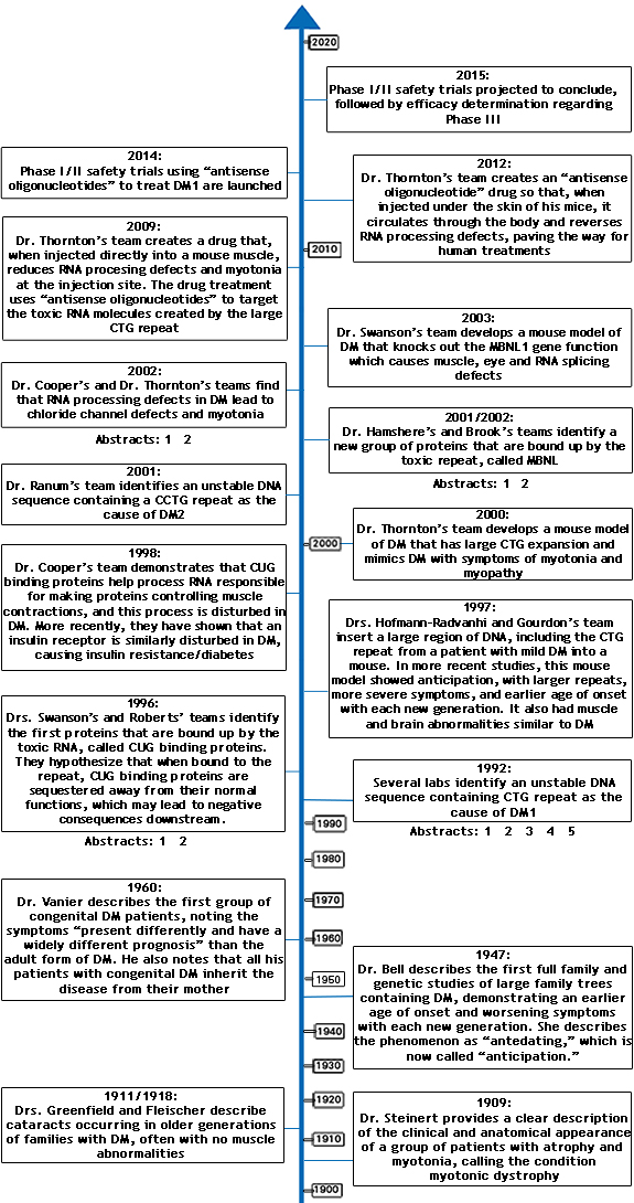
Timeline of key discoveries in myotonic dystrophy research since DM was first described in 1909. Click on the corresponding timeline entry for links to research abstracts or reviews. 

Abstracts not available for: Greenfield and Fleischer, 1911/1918; Bell, 1947; Vanier, 1960.


HTML Map

Impact of Childhood and Congenital DM on Quality of Life

Published on Tue, 06/25/2013

Nicholas Johnson, MD, and researchers at the University of Rochester recently published an article in The Journal of Child Neurology that describes the impact of childhood and congenital myotonic dystrophy on quality of life. The authors interviewed 21 children with childhood and congenital myotonic dystrophy and 13 parents. After recording these interviews, the authors reviewed transcripts to identify the most important symptoms to parents and children. Overall, participants reported 189 different symptoms. 

Dr. Johnson and his colleagues found that many of the parents and children identified trouble speaking (dysarthria) as the primary symptom that impacted the children’s lives. Other participants identified learning difficulties and problems concentrating as life altering symptoms. Although many of the study participants did not identify a diagnosis of autism specifically, autistic traits, such as a narrow scope of interest, repetitive speech, inappropriate social responses, and inflexibility were reported. 

As expected, many of the symptoms affecting those with congenital and childhood myotonic dystrophy are different from the symptoms of adult-onset myotonic dystrophy type-1. Prior work by Chad Heatwole, MD, MS-CI, the senior author on this study, identified fatigue, hand and finger weakness, and difficulty walking as significantly impacting the quality of life of those with adult-onset myotonic dystrophy type-1. While these symptoms were also reported in children with myotonic dystrophy, their presentation and significance were different. 

Importantly, many symptoms of congenital and childhood myotonic dystrophy, such as communication difficulties, already have available treatments. The authors hope that by identifying the wide range of symptoms affecting children with myotonic dystrophy, doctors will be able to identify critical symptoms earlier and initiate timely treatment strategies.

Dr. Johnson and University of Rochester researchers, with the support of MDF, have used information from this study to develop a survey, which was distributed to US, Canadian and Swedish patients with congenital and childhood myotonic dystrophy. Results from this international group of patients will be used to further define and prioritize the most important symptoms to patients with congenital and childhood myotonic dystrophy. Ultimately this data will be used to help guide researchers in designing future therapeutic trials for these populations. 

06/25/2013

A New Study Provides Hope for DM Treatments

Published on Tue, 04/16/2013

Maurice Swanson, Ph.D., Professor of Molecular Genetics and Microbiology at University of Florida, Gainesville, and a team of researchers have found that the muscleblind-like 2 (MBNL2) protein in the central nervous system (CNS) may be responsible for the neurological impacts of myotonic dystrophy (DM), providing hope for new treatments. Muscleblind is a type of protein that plays an important role in switching proteins typically found only in babies to proteins found in adults. If this switch isn’t made, an imbalance exists that leads to myotonic dystrophy.

Dr. Swanson states that the team’s work seeks to understand what causes myotonic dystrophy beyond the mutations in the DM1 and DM2 genes.

Dr. Swanson examined which genes were affected by loss of MBNL2 in the brain and found more than 800 affected genes. Many of them had one thing in common: the encoded protein could be made in both fetal and adult forms and MBNL2 appeared to regulate which version was created, according to an article in Neurology Today. One persistent concern that people living with DM1 and DM2 have is the effects of this disease on the brain. “People who don’t have DM usually feel refreshed after a night’s sleep. Myotonic dystrophy patients do not routinely achieve a normal sleep pattern; instead, they have an interrupted series of sleep-wake patterns that do not allow for deep, restful sleep cycles”.

Dr. Swanson created a mouse that lacks the MBNL2 protein as an animal model for DM effects on the CNS. These mice showed normal skeletal muscle structure and function. However, the mice did have DM-related sleep issues, such as a higher number of REM sleep episodes and more REM sleep in general, leading to less restful sleep. In mice lacking MBNL1, another member of the MBNL protein family, the skeletal muscle effects were similar to what is seen in DM. But the central nervous system was not affected, according to Dr. Swanson. 

“What we would like to do now is identify the specific cellular events that are abnormal in the DM brain and see if there is something we can do to treat these disease manifestations with focused therapy development. We would also like to understand the heart and muscle problems in DM. We have developed mice with DM-associated problems and we want to use these mouse models to develop effective drug treatments. Also, we want to understand what is so different about the congenital form of DM. Why does it manifest in babies and children? If we can develop animal models for congenital DM, then we can begin to address the important question of what goes wrong during fetal life,” explains Dr. Swanson.

Recently, therapy development for DM has accelerated and treatments based on anti-sense oligonucleotides will hopefully enter clinical trials in the near future. These new studies focused on the roles of MBNL proteins in CNS function should lead to alternative therapeutic strategies designed to reverse effects caused by expression of the mutant DM1 and DM2 genes.

04/16/2013

Ritalin Could Curb Excessive Daytime Sleepiness

Published on Wed, 01/23/2013

Methylphenidate, a psycho-stimulant drug, also known by its 1948 trademarked name of Ritalin, could be useful in the treatment of excessive daytime sleepiness (EDS) for DM1 patients, according to a recent study conducted by The Department of Human Genetics at the Centre Hospitalier Universitaire de Quebec in Quebec City, Canada.  

A total of 24 French-Canadian patients who had DM1 and an Epworth Sleepiness Scale score of more than 10 (0-9: normal, 10-24: sleepy and medical advice should be sought) were invited to participate in the 3-week crossover trial of 20-mg/d of methylphenidate versus a placebo. There were two groups in the study; one group took the drug for 3 weeks, followed by a 2-week washout period, followed by 3 weeks of placebo, and the other group took placebo for 3 weeks, followed by a 2-week washout period, followed by 3 weeks of drug. Of the 24 patients (12 men; 12 women; median age 46 years), 17 completed the study. 

Patients’ Daytime Sleepiness Scale and the Epworth Sleepiness Scale, measured at the end of each 3-week period, were used to measure the effectiveness of methylphenidate as a treatment for EDS. The drug’s effectiveness was also determined by a mean sleep latency test, patients’ energy and vitality after the study, and patients’ moods. Tolerability to treatment was monitored by blood pressure, the results of an echocardiogram, and other lab tests. Adverse reactions to the treatment were based on patients’ reporting and were recorded at each visit to the clinic. 

The study concluded that a single 20-mg/d dose of methylphenidate significantly reduced daytime sleepiness in this small group of patients with DM1. The median scores on the Epworth Sleepiness Scale and the Daytime Sleepiness Scale showed a significant change. However, measurements of patients’ moods, energy, and vitality showed no changes, and the mean sleep latency test showed no significant changes. The most common adverse effects included nausea, loss of appetite, and palpitations, as reported by more patients who were treated with methylphenidate than by those receiving the placebo. Three patients stopped taking methylphenidate due to adverse effects that arose during treatment, which included diarrhea, nervousness and irritability. One patient died during the trial, but the autopsy results eliminated methylphenidate as the cause of death. 

For more information about this study, click here
 

01/23/2013

MDF Awards $200,000 for Research into Treatments for DM

Published on Wed, 01/23/2013

MENLO PARK, CA (January 2013): MDF is pleased to announce its most recent round of Fund-A-Fellow postdoctoral fellowship research grant awards. In January 2013, MDF awarded two $100,000 grants to postdoctoral Fellows working in universities and research facilities to encourage basic research in the management, treatment and cure of myotonic dystrophy (DM) for a commitment of $200,000. 

This award cycle brings the total research funding awarded by MDF to over $1.5M since its founding in 2006, and builds on MDF’s commitment to increasing the number of investigators focused on myotonic dystrophy research.  To date, MDF Fellows have attracted additional research funding from larger organizations such as the National Institutes of Health, have helped influence interest in DM research at major pharmaceutical companies, and have risen to senior positions at academic and clinical settings across the United States. MDF Fund-a-Fellows have collaboratively deepened understanding of and commitment to myotonic dystrophy research significantly since the program’s founding in 2009.

Each of the two 2013-2014 FAF recipients will receive $50,000 a year for two years. The applicant pool for this year’s grant awards was extremely competitive and totaled 15, the largest ever for MDF fellowship funding.  Applications were reviewed by a panel of distinguished international researchers and clinicians in the field of myotonic dystrophy. Final selections were determined by the MDF Board of Directors. 

The two 2013-2014 $100,000 postdoctoral fellowship grants have been awarded to: Dr. Suzanne Rzuczek, Ph.D., The Scripps Research Institute – Florida; and Dr. Ayal Hendel, Ph.D., Stanford School of Medicine.  

Research Information

Dr. Ayal Hendel, Ph.D.

CTG/CAG repeat tracts represent the genetic basis for more than a dozen inherited dominant neurological disorders including Myotonic Dystrophy type 1 (DM1) and Huntington’s disease that currently have no cure.  Despite the multitude pathologies underlying these devastating disorders, they all share common etiology: the expansion of CTG/CAG repeats. Interestingly, expanded CTG/CAG repeats have been shown to be prone to double-strand breaks (DSBs).  Moreover, it was shown that the repair of the DSBs leads mainly to repeat contractions. These findings suggest that controlled DSBs might provide a way to induce repeat contractions that will correct disease-causing mutation and reduce disease risk. Dr. Hendel’s research will harness a unique genome editing technology in combination with induced pluripotent stem cells to examine the contribution of DSB repair to the stimulation of repeat contractions in DM1 cells, ultimately exploring new cellular and molecular DM1 pathological mechanisms involved in myotonic dystrophy.

Dr. Suzanne Rzuczek, Ph.D.

Dr. Rzuzcek, in conjunction with the Disney Lab of The Scripps Institute, plans on using small molecules to disrupt the interaction between repeating CUG RNA and MBNL1 (muscleblind-like 1 protein), freeing it to function normally. The Disney Lab, of which Dr. Rzuczek is a member, has designed and synthesized several compounds that specifically bind the expanded CUG RNA and disrupt the interaction with MBNL1 in vitro. These compounds contain CUG-binding small molecules tethered together by a spacer. This approach is called modular assembly. Recently the spacer has been optimized to increase bioactivity and cell uptake. Dr. Rzuczek and her colleagues will improve the activity of the modularly assembled compounds using two approaches. The first will use the repeating DM1 RNA as a template to assemble small compounds with their optimized spacer into large modularly assembled structures within cells. This process is known as in situ click chemistry. The second approach will screen many small molecules that are similar to the known binders of DM1 RNA. Hit compounds will be screened in cells using a method that detects low levels of bioactivity. Compounds that are active in cells will be modularly assembled on the Disney-optimized spacer to enhance the interaction with DM1 RNA and improve bioactivity. If successful, these compounds could become a treatment for myotonic dystrophy. 


01/23/2013

Mexiletine for Myotonia: A New Use for an Old Heart Drug?

Published on Tue, 10/23/2012

A generic cardiovascular drug called mexiletine, initially developed to treat heart rhythm abnormalities, appears to hold some potential for treating muscle stiffness and other symptoms of non-dystrophic myotonias (NDMs), a rare group of disorders without progressive muscle wasting and weakness, that have abnormalities in the function of ion channels in the muscle cells (chloride or sodium channels).  The abnormal ion channel function leads to stiffness and delayed relaxation of muscle following grip or tight closing of the eyes (myotonia).  Myotonic dystrophy (DM) has as one of its alterations abnormal function of the chloride channel.  This causes myotonia and stiffness, and, like the patients with non-dystrophic myotonia, DM patients show a beneficial response to treatment with mexiletine.

A study published in the Journal of the American Medical Association (JAMA) in early October evaluated the drug in 59 people with NDMs. The study, sponsored by the National Institutes of Health’s Rare Disease Clinical Research Network, randomized patients with one of the rare forms of non-dystrophic myotonia to either the study drug (one capsule of mexiletine three times a day for four weeks) or placebo. After four weeks, patients stopped taking either for a week then switched to the opposite treatment for four weeks. 
 
When patients took the study drug, their muscle stiffness improved up to 40 percent and their pain scores fell by 17%. Objective tests also found that mexiletine reduced abnormal electrical activity (myotonic discharges) in the muscles that led to the spasms and stiffness. Patients also experienced significant improvements in their overall quality of life. The most common side effects were gastrointestinal, such as diarrhea, nausea, and heartburn. Click here to read an abstract of the article.
 
This study echoes findings on mexiletine and its beneficial effects on myotonia published in 2010 by Drs. Moxley, Logigian, Martens, Thornton et al.  The 2010 study, conducted at the University of Rochester with a group of 20 DM1 patients, found that grip relaxation time improved by 50% and muscle stiffness decreased when patients took mexiletine compared to placebo. Click here to read an abstract on the 2010 Rochester study.
 
Mexiletine works by slowing the rate of movement of sodium ions into muscle cells, reducing or eliminating the tendency of the muscle to maintain a prolonged contraction.  This enables the muscle cell to relax properly and be ready for the next contraction.  Mexiletine works for several hours and it needs to be taken two or three times daily to exert continued beneficial effects.
 
The drug is not approved for treating either NDM or DM, but doctors can prescribe it “off label.” However, as with any drug, there are risks and benefits and only your doctor, with your input, can decide if it might work for you.
 
10/23/2012

Dysregulation of Circular RNAs in DM1

Published on Thu, 05/17/2012

Circular RNA Primer

Cells contain a striking diversity of RNA types, many of which have been implicated in the pathogenesis of neuromuscular disease. Unlike most RNAs, circular RNAs (circRNAs) are single-stranded, covalently closed loops. CircRNAs arise via one of three mechanisms: (a) direct ligation of 5′ and 3′ ends of linear RNAs, (b) as intermediates in RNA processing reactions, or (c) via “back splicing,” when a downstream 5′ splice site (donor) is joined to an upstream 3′ splice site (acceptor). A variety of biologic roles for circRNAs have been identified.

Presence of circRNA in DM1 Skeletal Muscle

While aberrant RNA splicing represents a central disease mechanism in DM1, virtually nothing is known regarding the potential for dysregulation of circRNAs. Dr. Fabio Martelli (IRCCS Policlinico San Donato) and colleagues have recently published an analysis of dysregulation of circRNAs in DM1 patient skeletal muscle biopsies (Voellenkle et al., 2019). The authors show that specific myogenesis-associated circRNAs are altered in DM1 biopsies and in DM1 patient myogenic cell cultures.

The research team identified specific circRNAs through review of 30 published DM1 RNAseq databases—relative abundance of a circRNA to its linear counterpart was used as an initial filter, followed by comparison to a list of circRNAs that were previously identified in human or murine myoblasts. Thus, the analysis was not comprehensive, but geared toward identification of transcripts most likely to be dysregulated in skeletal muscle tissue. Candidate circRNAs meeting the investigators’ criteria then were validated using qPCR of skeletal muscles from DM1 subjects and age-/sex-matched controls. Primer specificity was confirmed and the possibility that results were due to a general increase in transcription in DM1 was excluded, and results were confirmed in independent muscle biopsies.

Taken together, four circRNAs—circCDYL, circHIPK3, circRTN4_03, and circZNF609—exhibited significantly increased circular-to-linear RNA ratio in DM1 muscles versus controls. The research team subsequently used receiver operating characteristic curve analysis and confirmed that a transcript’s circular-to-linear ratio could discriminate between DM1 and healthy controls. Finally, the increase in circular fraction for the four circRNAs correlated with a variety of clinical and molecular characteristics of study subjects. Circular fraction ratios correlated with both skeletal muscle strength and splicing biomarkers of disease severity.  Moreover, circular fraction was higher in the more severely affected DM1 patients. Induction of two of the dysregulated circRNAs (circCDYL and circRTN4) was also detected in plasma. Finally, analyses of DM1 myogenic cell lines identified a pattern of circRNA dysregulation that was, in part, similar to data obtained in patient muscle biopsies.

Potential Utility of Dysregulated circRNAs in DM1

The research team self-identified caveats and described these findings as pilot data. Any putative contributing role that dysregulated circRNAs may have in the pathogenesis of DM1 is currently unknown. Yet the discovery of specific, dysregulated circRNAs in DM1 skeletal muscle, if confirmed, may offer advantages for drug development efforts—circRNAs are exceptionally stable in that they are resistant to exonuclease degradation and their dysregulation was detectable in plasma and myogenic cell lines from DM1 patients. These traits make them amenable to use as pharmacodynamic biomarkers for clinical studies and trials in DM1.

Reference:

Dysregulation of Circular RNAs in Myotonic Dystrophy Type 1.
Voellenkle C, Perfetti A, Carrara M, Fuschi P, Renna LV, Longo M, Sain SB, Cardani R, Valaperta R, Silvestri G, Legnini I, Bozzoni I, Furling D, Gaetano C, Falcone G, Meola G, Martelli F.
Int J Mol Sci. 2019 Apr 19;20(8). pii: E1938. doi: 10.3390/ijms20081938.

What is the Actual Progenitor Allele Length in Any Given DM1 Patient?

Published on Thu, 05/17/2012

The Elusive Quantification of Repeat Length

In a disease that exhibits somatic mosaicism, somatic cell instability, and the consequent tissue-to-tissue variability in expanded repeat length, assigning an “actual progenitor repeat length” value to individual DM1 patients has been problematic. The connotations here are obvious—how do we use repeat length for essential drug development functions from molecular biomarkers to genotype-phenotype analyses to stratification of patients in interventional clinical trials, if the parameter is hard to pin down? A recent paper from a multi-site team (Universidad de Costa Rica, University of Texas MD Anderson Cancer Center, and University of Glasgow) attempts to determine the optimal body fluid/tissue to sample and thereby yield insight into the best path forward for clinical studies and interventional clinical trials (Corrales et al., 2019).

Saliva as an Accessible and Reliable Source for DM1 Mutation Testing

Dr. Fernando Morales and colleagues sought to build on their prior findings (Morales et al., 2012 & 2016) that used small pool-PCR (SP-PCR) to control for somatic instability in estimations of progenitor allele length measured in blood. The goal was to improve upon allele length correlations with age of DM1 onset. In their latest work, the research team reports out on comparison of saliva vs. blood as the analyte source for progenitor allele length determinations.

This report was based upon analysis of progenitor allele length in saliva and blood from 40 DM1 patients that had been characterized for age of onset; screening also assessed for presence of variant repeats and methylation of CTCF binding sites adjacent to the DMPK locus, as these may be modifiers of somatic instability. Modal allele length was slightly larger in saliva (529 repeats) than concurrently collected blood samples (486 repeats). Progenitor allele length then was estimated as the lower boundary of allele distribution from SP-PCR—values from the two sample sources were highly correlated and, again, were higher in saliva than blood (414 vs. 310).

Analyses showed that progenitor allele length estimated from blood samples explained 75% of the variation in DM1 age of onset, while that from saliva explained 66% of the variation. The authors suggest that the “true progenitor allele length” needed for genotype-phenotype studies and other preclinical and clinical development purposes is more likely reflected by the values obtained from blood samples.

Additional single molecule SP-PCR studies, excluding two CDM cases, revealed greater somatic instability in blood than in saliva. The research team also showed that the lower boundary of allele distribution was slightly higher in saliva than in blood, while the overall degree of somatic variation was typically lower in saliva than in blood. Finally, analyses of repeat variants and methylation levels as putative modifiers of somatic instability showed that neither were significant factors.

Blood or Saliva?

The authors of this paper summarize the compelling literature case against use of skeletal muscle samples (essentially the confounding effect of tissue-specific rate of somatic expansion) to estimate progenitor allele size, bringing the choice down to blood or saliva. These data show that somatic mosaicism is comparable in blood and saliva DNA from DM1 patients, while saliva is obtained by considerably less invasive means—a feature that is potentially vital for interventional clinical trials in CDM or repeated sampling to assess efficacy of a candidate therapeutic in either CDM or DM1.

References:

Analysis of mutational dynamics at the DMPK (CTG)n locus identifies saliva as a suitable DNA sample source for genetic analysis in myotonic dystrophy type 1.
Corrales E, Vásquez M, Zhang B, Santamaría-Ulloa C, Cuenca P, Krahe R, Monckton DG, Morales F.
PLoS One. 2019 May 2;14(5):e0216407. doi: 10.1371/journal.pone.0216407. eCollection 2019.

Somatic instability of the expanded CTG triplet repeat in myotonic dystrophy type 1 is a heritable quantitative trait and modifier of disease severity.
Morales F, Couto JM, Higham CF, Hogg G, Cuenca P, Braida C, Wilson RH, Adam B, del Valle G, Brian R, Sittenfeld M, Ashizawa T, Wilcox A, Wilcox DE, Monckton DG.
Hum Mol Genet. 2012 Aug 15;21(16):3558-67. doi: 10.1093/hmg/dds185. Epub 2012 May 16.

A polymorphism in the MSH3 mismatch repair gene is associated with the levels of somatic instability of the expanded CTG repeat in the blood DNA of myotonic dystrophy type 1 patients.
Morales F, Vásquez M, Santamaría C, Cuenca P, Corrales E, Monckton DG.
DNA Repair (Amst). 2016 Apr;40:57-66. doi: 10.1016/j.dnarep.2016.01.001. Epub 2016 Mar 8.