News  Donate!

Research

Using iPSC-Derived Cardiomyocytes to Understand DM1

Published on Tue, 04/10/2018

Induced pluripotential stem cell (iPSC) technology has provided opportunities to better understand disease mechanisms as well as to facilitate drug discovery and development programs. With the attendant molecular, cellular, and disease backgrounds of readily expandable, patient-derived cells, iPSC “disease-in-a-dish” models have supported the translation of candidate therapeutics from discovery into clinical testing.

As long as limitations are recognized, such as potential genetic and epigenetic divergence from primary cells, iPSC modeling of myotonic dystrophy (DM) may be essential to mechanistic understanding and development of treatments for the disease, particular when patient tissues are difficult to impossible to access (e.g., CNS and heart). MDF has facilitated the development of human DM1 iPSC lines, now available through RUCDR Infinite Biologics, with DM2 lines to follow.

Development of iPSC-Derived Cardiomyocytes

Reported in a newly published study, Dr. Federica Sangiuolo (University of Rome Tor Vergata) and colleagues have developed iPSC lines from two DM1 patients and two healthy controls. They characterized cardiomyocytes derived from these lines, with the goal of determining whether they could recapitulate at least some of the major traits of the DM1 heart. Myocyte characterization included RT-qPCR to quantify RNA expression, FISH and IF for nuclear foci, RT-PCR for splice variants, whole-cell patch clamp to characterize cardiomyocyte electrophysiology, and atomic force microscopy (AFM) to characterize biomechanical traits.

Traits of iPSC-Derived Cardiomyocytes—DM1 versus Wild Type

Most iPSC-derived cardiomyocytes exhibited a ventricular-like phenotype. DM1-derived cardiomyocytes contrasted with wild type controls in expressing nuclear foci and mis-slicing of each transcript evaluated (MBNL1, MBNL2, TNNT2 and SCN5A); DM1 derivatives also showed down-regulation of key cardiac ion channel transcripts (CACNA1C, KCNH2, KCNQ1, KCND3, and SCH5A) compared with controls. The research team noted novel differences in nuclear morphology in DM1 derivatives, potentially related to altered expression of lamin A.

Whole-cell patch clamp recordings of disaggregated single cardiomyocytes showed an abnormal electrophysiological profile only in the DM1 derivatives. DM1 ventricular myocyte-like derivatives exhibited lower spontaneous action potential rates, lower peak amplitude, and longer time to reach peak from threshold. Electrophysiologic parameters of DM1 cardiomyocytes could be altered by drugs targeting cardiac ion channels.

Using AFM, the research team identified differences in beat frequency and synchronicity of beats in DM1 iPSC-derived cardiomyocytes—describing the overall pattern as indicative of instability, with reduced frequency and the appearance of non-synchronous oscillation patterns when compared to wild type. The predominance of ventricular-like myocytes, and relative absence of nodal or other conduction system-related cardiomyocytes, may make it more difficult for iPSC-based models to be informative of the conduction system-specific abnormalities known to characterize DM1.

Overall, DM1 iPSC-derived cardiomyocytes recapitulated the main morphologic and molecular markers of the disease, including CTG expansions, nuclear foci, splicopathy of the mRNAs evaluated, and altered expression of several known cardiac ion channels. Alterations in electrophysiological parameters and biomechanical behavior were interpreted as consistent with unstable beating.

Reference:

Modelling the pathogenesis of Myotonic Dystrophy type 1 cardiac phenotype through human iPSC-derived cardiomyocytes.
Spitalieri P, Talarico RV, Caioli S, Murdocca M, Serafino A, Girasole M, Dinarelli S, Longo G, Pucci S, Botta A, Novelli G, Zona C, Mango R, Sangiuolo F.
J Mol Cell Cardiol. 2018 Mar 15. pii: S0022-2828(18)30083-X. doi: 10.1016/j.yjmcc.2018.03.012. [Epub ahead of print]

Regulation of MBNL1 Localization and Function in the CNS

Published on Mon, 04/09/2018

Role of Muscleblind Proteins in Health and Disease

Muscleblind (MBNL) proteins play a central role in the pathogenesis of myotonic dystrophy (DM), as MBNL depletion below a threshold level triggers a range of mRNA splicing, polyadenylation, localization, and processing defects and, thereby, the production of temporally inappropriate protein isoforms. This compromise of function for a wide range of proteins, in turn, leads to the multi-organ system phenotypes that characterize DM. To understand MBNL’s roles in normal development and function, and dysfunction in DM, it is essential to elucidate mechanisms underlying its spatial and temporal localization and steps necessary for its functional activation/inactivation.

Dissecting the Role of MBNL in the CNS

Understanding the regulation and functional roles of MBNL proteins in the CNS represents a critically important problem in determining pathogenic mechanisms underlying the nervous system phenotype in DM. Dr. Guey-Shin Wang at National Yang-Ming University and Academia Sinica (Taiwan) have published an article in Cell Reports that MBNL1 localized to the cytoplasm, but not that retained in the nucleus, plays a key role in neurite morphogenesis and show that this role is disrupted in DM1.

Using FLAG-tagged isoforms of MBNL1 (with and without exon 5) expressed in hippocampal neuron cultures, Dr. Wang and colleagues demonstrated that MBNL1 that included exon 5 was retained in the nucleus and had no impact on neuronal development, while exon 5-deleted MBNL1, which translocates to the cytoplasm, enhanced dendrite and axon morphogenesis.

Consistent with this role for MBNL1 in regulating neurite differentiation and the differentiation failure in DM1, the research team showed that either MBNL1 knockdown or overexpression of 960-repeat DMPK mRNA (DMPK-CUG960) resulted in delayed neurite maturation. Moreover, the cytoplasmic (exon 5 deleted) MBNL1 isoform, but not the nuclear-localized isoform, was found to rescue maturation defects in neurons expressing DMPK-CUG960 in a dose-dependent manner.

Mechanisms Regulating Neuronal Subcellular Distribution of MBNL1

Analysis of the developmental distribution of the two MBNL1 isoforms led to the puzzling finding that nuclear or cytoplasmic localization was independent of exon 5 inclusion/exclusion, suggesting that another factor regulated MBNL1 localization in the developing brain. The research team then demonstrated that Lys 63-linked ubiquitination of MBNL1 and neuronal activity level regulated its cellular localization. Finally, it was shown that deubiquitination caused by expanded repeat mRNA resulted in a cytoplasmic-to-nuclear translocation of MBNL1 and corresponding alterations in neurite morphogenesis—these consequences were prevented by deubiquitination inhibitors.

Taken together, Dr. Wang and colleagues have shown that MBNL1 plays a key role in neuronal differentiation and that disruption of its cellular localization alters functionally significant steps in neuronal structure and function that likely underlie the CNS consequences of DM. Moreover, their identification of the regulatory role that the ubiquitination/deubiquitination status plays in the cytoplasmic localization of MBNL1 suggests a novel potential target for therapeutic development in DM1.

Reference:

Ubiquitination of MBNL1 Is Required for Its Cytoplasmic Localization and Function in Promoting Neurite Outgrowth.
Wang PY, Chang KT, Lin YM, Kuo TY, Wang GS.
Cell Rep. 2018 Feb 27;22(9):2294-2306. doi: 10.1016/j.celrep.2018.02.025.

NHGRI Genomic Medicine Research Initiative

Published on Mon, 04/09/2018

The National Human Genome Research Institute (NHGRI) has issued two new Funding Opportunity Announcements in support of research to utilize a patient’s own genomic information to inform clinical care and health outcomes. These Announcements represent a potential opportunity to apply an understanding of the genomics of those living with myotonic dystrophy (DM) to clinical management and treatment settings.

The stated objective of the NHGRI program is to “stimulate innovation and advance our understanding of when, where and how best to implement the use of genomic information and technologies in clinical care in all populations.” The program utilizes both R01 (PAR-18-735) and R21 (PAR-18-736) grant mechanisms. R01 applications are limited to $500,000 direct costs/year and 4 years; R21 applications to $275,000 total direct costs over 2 years. The PAR designation implies that applications will be assigned to a dedicated study section (Special Emphasis Panel or other) for review; there are no set-aside funds designated for this initiative.

Eligible activities include, but are not limited to, research studies on genome sequencing for disease diagnosis, treatment, or healthcare utilization. Interested applicants should see the Program Announcements referenced above and are strongly encouraged to discuss plans for applications with the Program Director identified in the Announcements.

Could Myotonic Dystrophy Be Included in Newborn Screening? Experts Say It’s Time to Explore

Published on Wed, 07/23/2025

Bringing Myotonic Dystrophy into the Newborn Screening Conversation

On February 14, 2025, the RNA Institute and the Myotonic Dystrophy Foundation (MDF) organized and co-hosted the first ever symposium exploring how to include myotonic dystrophy (DM) in newborn screening. Newborn screening is one of the most successful public health services testing all babies shortly after birth for certain genetic, life-threatening diseases. Conditions included in newborn screening are serious and life threatening and without early diagnosis would result in irreparable harm and death. Conditions included in newborn screening are also diseases that do not have treatments but benefit from early disease management thereby significantly improving clinical outcomes. Newborn screening identifies babies at risk for these diseases before symptoms start, ensures the best possible outcomes for all babies, and limits pain and suffering.

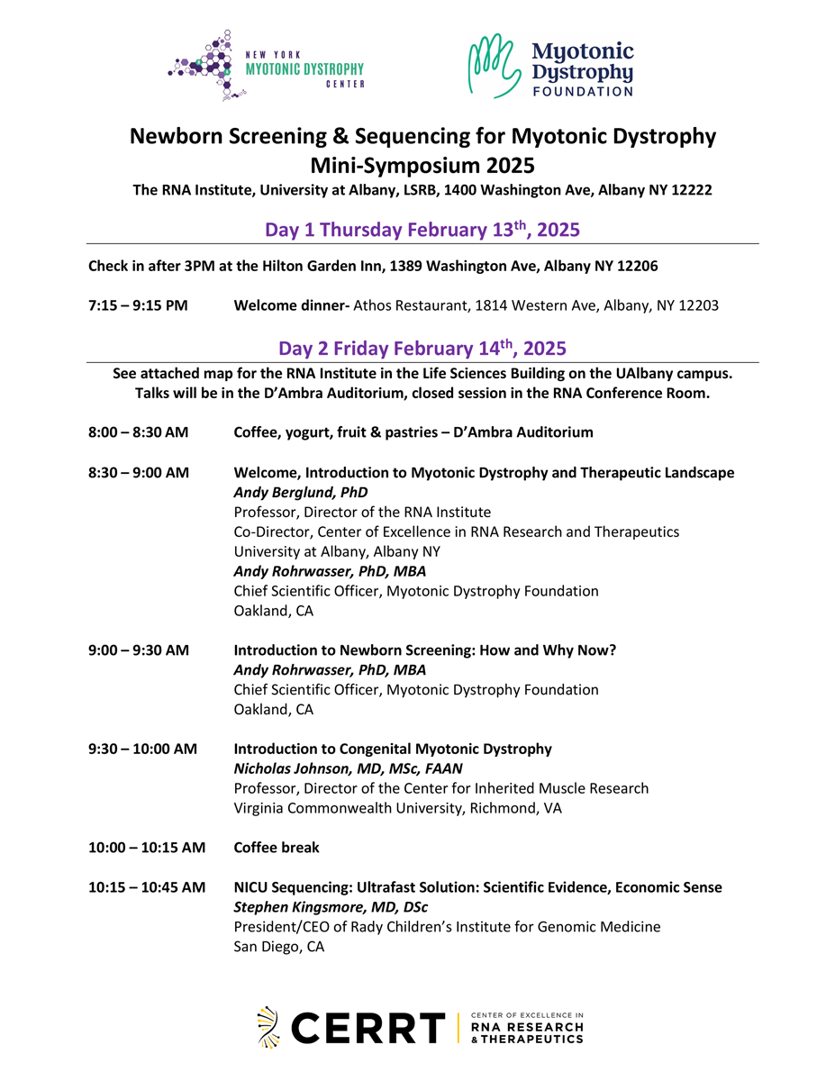
Click here to download the agenda from the Newborn Screening & Sequencing for Myotonic Dystrophy Mini-Symposium 2025! >>>

Looking for more detail? Scroll down for scientific and expert-level summaries, including a Scientific Abstract and a deeper technical overview.

The Power and Promise of Genome Sequencing

The meeting brought together researchers, doctors, public health leaders, industry experts, and patient advocates to share progress in genome sequencing, diagnostic tools, and pilot newborn screening programs. Laboratory tests using genome sequencing allow the identification of a select set of genetic diseases based on changes in a person’s DNA. Diagnostic genome sequencing is a comprehensive genetic test performed in a specialized laboratory. The test analyzes an individual's entire genome or DNA to identify potential disease-causing changes. It is increasingly used to diagnose rare and inherited diseases, particularly when other tests are either inconclusive or simply not available. By examining the entire genome, the test can detect changes that might be missed by other tests, potentially leading to a faster and more accurate diagnosis. Although such tests could help diagnose babies faster and cut healthcare costs, obstacles for implementing the tests include high costs, limited state funding, and the lack of treatments for infants with DM. Experts also discussed ethical concerns—like testing infants for untreatable conditions—but noted that early detection of DM would allow for planning, including engaging in preventive and pre-symptomatic health measures such as regular cardiac monitoring, raise awareness of the serious impact of anesthesia, and would also support research.

Next Steps Toward a Future with Newborn Screening for DM

Participants recommended the following next steps: tracking DM patients over time, piloting health-record studies to identify patients with DM earlier, promoting testing and reimbursement policies, educating medical providers about genome sequencing-based testing and early diagnostics through newborn screening, exploring and advocating for the addition of DM in prenatal screens, and involving the community to build supporting data and resources needed for DM newborn screening in the future.

Thank You to Our Hosts and Organizers


Scientific Summary: Newborn Screening for Myotonic Dystrophy

Background: On February 14, 2025, the RNA Institute and the Myotonic Dystrophy Foundation held a mini symposium to discuss how myotonic dystrophy (DM) could be added to newborn screening. Newborn screening is one of the most successful public health services testing all babies shortly after birth for certain life-threatening diseases. Conditions included in newborn screening are serious and life threatening and without early diagnosis would result in irreparable harm and death. Newborn screening identifies babies at risk before symptoms start, ensures best possible outcomes for all babies, and limits pain and suffering. Methods: The meeting brought together researchers, clinicians, public-health leaders, industry experts, and patient advocates. They reviewed advances in genome sequencing, diagnostic methods, and early pilot newborn screening programs.

Results: Experts agreed that rapid genome testing could speed up diagnosis and help lower healthcare costs. However, several barriers were raised:

  • High testing costs
  • Limited state funding and a shortage of infrastructure and trained personnel performing genome sequencing based newborn screening
  • No existing treatments for infants with DM

Ethical Discussion: Participants debated the ethics of testing newborns for still untreatable conditions. Despite these concerns, they noted that early detection helps parents prepare and supports research progress. Recommendations: The group proposed several action items:

  1. Follow diagnosed infants over time
  2. Conduct pilot studies using electronic health records to identify undiagnosed patients
  3. Advocate for supportive policies and pilot studies ultimately enabling newborn screening for DM
  4. Educate healthcare providers about DM screening and genetic testing solutions
  5. Explore adding DM to prenatal screen panels
  6. Engage families and communities to build support for the adoption of newborn screening for DM

Conclusion: While acknowledging financial, logistical, and ethical challenges, the symposium concluded that early detection of DM offers clear benefits. A coordinated strategy—combining research, policy advocacy, provider education, and community engagement—is essential for future implementation.


Full Technical Summary: Symposium on Genomic Sequencing and Myotonic Dystrophy

Summary of Newborn Screening & Sequencing for Myotonic Dystrophy

Newborn screening (NBS) is a vital and extremely successful public health program aimed at the early identification of conditions that can affect a child’s long-term health or survival. Early detection, diagnosis, and intervention – commonly before the onset of symptoms - can prevent death or disability, alleviate physical, emotional, and economic suffering, and enable children to reach their full potential. With the advancement of genomic sequencing technologies, there is growing interest and opportunity in expanding NBS to include a broader range of genetic disorders. This broadened scope includes conditions, like myotonic dystrophy (DM), for which early treatment may not yet be available but where early diagnosis can inform family planning, clinical monitoring, and guide future therapeutic development. The following summary synthesizes the discussion from a recent Myotonic Dystrophy mini-symposium hosted by the University at Albany’s RNA Institute and co-organized by the Myotonic Dystrophy Foundation and the RNA Institute. The following notes highlight the current state of the field, challenges, and future directions of NBS and genetic testing for DM.

Key Players weighed in on Newborn Screening

The discussions involved a diverse group of stakeholders from academia, industry, public health, and patient advocacy groups. Andy Berglund, PhD (Director, RNA Institute & Chair of Myotonic Dystrophy Foundation Scientific Advisory Committee) and Andy Rohrwasser, PhD, MBA (Chief Scientific Officer, Myotonic Dystrophy Foundation) co-organized and facilitated the meeting with key stakeholders in the field.

In a public session open to the community, key invited experts and opinion leaders summarized critical insights into NBS, myotonic dystrophy (DM), how NBS could be applied to DM, clinical and economic benefits supporting genomics based newborn screening, as well as technology opportunities today that could bridge until genomic NBS solutions would become available.

  • Introduction to Myotonic Dystrophy and Therapeutic Landscape; Andy Berglund, PhD; Professor, Director of the RNA Institute, Co-Director, Center of Excellence in RNA Research and Therapeutics, University at Albany, Albany NY
  • Introduction to Newborn Screening: How and Why Now? Andy Rohrwasser, PhD, MBA, Chief Scientific Officer, Myotonic Dystrophy Foundation, Oakland, CA
  • Introduction to Congenital Myotonic Dystrophy, Nicholas Johnson, MD, MSc, FAAN, Professor, Director of the Center for Inherited Muscle Research, Virginia Commonwealth University, Richmond, VA
  • NICU Sequencing: Ultrafast Solution: Scientific Evidence, Economic Sense, Stephen Kingsmore, MD, DSc; President/CEO of Rady Children’s Institute for Genomic Medicine, San Diego, CA
  • Pilot Study: Towards Population Wide Sequencing; Wendy Chung, MD, PhD; Chief of Pediatrics, Boston Children’s Hospital, Professor, Harvard Medical School, Boston MA
  • Newborn Sequencing in Public Health: How would it work in NY? Michele Caggana, ScD, FACMG; Deputy Director, Division of Genetics; Director, Newborn Screening Program; Department of Health, Wadsworth Center, Albany NY
  • Rapid Advance in Analysis and Interpretation; Opportunities in EHR mining - before population wide screening? Mark Yandell, PhD; Professor, Director Eccles Institute Bioinformatics Program; Technical Director, Utah Genome Project; The University of Utah, Salt Lake City, UT

Key Meeting Outcomes and Insights

Following general presentations to the public and interested scientific community members at the University at Albany, the key stakeholders held an in-person discussion with a few virtual attendees. Several important outcomes and insights emerged from the discussion. The Guardian Study was highlighted, as a large-scale genomic screening initiative in New York City, which has screened over 15,000 participants to date and reported a 3.3% positive screen rate with an average turnaround time of approximately 22 days.

Whole genome sequencing (WGS) in neonatal intensive care units (NICUs) was highlighted as the ultimate promise in the ultra-rapid diagnosis reducing time-to-diagnosis and eliminating diagnostic odysseys, but also reducing pain and suffering based on misdiagnoses and associated unnecessary diseases management, while also significantly reducing healthcare costs. The stakeholders also highlighted that repeat expansion disease testing is gaining attention, particularly for conditions such as congenital myotonic dystrophy.

While support for the need for increased testing was strong amongst the group, significant policy and infrastructure challenges were noted. Many states currently lack formal policies for reimbursement for WGS diagnostics, posing a significant fiscal challenge to NBS screening programs. While New York State has pilot programs, there is no statewide policy in place for the selection of disease candidates. Sequencing costs, estimated at around $60 per newborn, along with the need for equipment redundancy and trained personnel, were also raised as significant logistical barriers. The rigorous nature of the Recommended Uniform (newborn Screening) Panel (RUSP) nomination process, which involves a nine-month review process and a 120-day decision window requiring strong evidence of treatment efficacy and public health impact would be significant challenges at this time. This is further complicated by the current administration’s dissolution of the Secretary’s Advisory Committee for Heritable Disorders in Newborns and Children that halted the process and disrupted the mechanism for adding or removing disorders from the recommended panel.

The need for an effective treatment for newborns with DM was discussed as an important point needed to support NBS. Given that clinical trials are currently primarily focused on adults, it was noted that the timeline for approved treatments for newborns was unclear. Foundational to this lack of treatment however is long-term natural history and outcome data for children affected by the early onset form of DM.

Ethical and social considerations were also discussed by the group. Concerns were raised about the implications of screening for conditions without available treatments, including the significant psychological impacts to the affected families and issues associated with insurance and insurability. Clinicians and patient advocates raised the point that early diagnosis can significantly affect family planning and emotional well-being of DM families. There was universal recognition of need for further real-world data and comprehensive, long-term natural history studies, given that many symptomatic individuals remain undiagnosed.

Recommended Next Steps

Several next steps were recommended by the group to advance the field. In terms of research and data collection, more long-term natural history and outcome studies were recognized as essential to understand disease progression and optimal treatment timing. The latter was a key highlight for myotonic dystrophy type 1 (DM1), which is highly heterogeneous in presentation between affected individuals. Integrating real-world data and study data, exploring genotype-phenotype relationships through collaborations with organizations like MDF and academic networks were also raised as crucial next steps. Pilot studies, utilizing EHR mining identifying undiagnosed DM patients for example in the Intermountain Health system, could help assess the feasibility and outcomes of broader implementation and bridge towards the implementation of newborn screening.

From a policy and advocacy perspective, gaining provider buy-in is critical. Here, for example RTI-led focus groups and precedence from other diseases were identified to potentially help to identify provider perspectives and barriers as well as preparing comprehensive evidence packages for RUSP nominations, including cost-benefit analyses and patient impact data. While the likelihood of NBS screening for DM1 was recognized as being years away from reality, the potential of maternal screening initiatives for DM, potentially in partnership with professional organizations like American College of Obstetricians and Gynecologists (ACOG), American College of Medical Genetics and Genomics (ACMG) or Society for Maternal-Fetal Medicine (SMFM) and commercial reference laboratories through integration in existing maternal panels was recognized.

Community engagement was identified as a key focus area raised by the experts and stakeholders alike. The collective group believed that targeted educational materials should be developed to inform families about the opportunities, implications and options related to genetic testing. The involvement of a broad coalition of stakeholders, including families, advocacy groups, and clinicians, was felt to be essential to align priorities and address ethical considerations. The group also believed that current efforts should include ensuring equitable access to testing and support systems for newly diagnosed families.

In summary, while the implementation of NBS for DM faces significant logistics and regulatory hurdles given the lack of an approved treatment approach for newborns affected by the disease, there was recognition of the power, opportunity and necessity of screening and diagnostic sequencing to make an impact on the DM community.

Fixing DNA Typos: New Hope for Myotonic Dystrophy

Published on Wed, 07/23/2025

A recent publication in Nature Genetics explores base editing as a solution to repeat expansion diseases similar to myotonic dystrophy (DM). The summary below was created by Asmer Aliyeva, PhD Candidate in Dr. Andy Berglund’s lab at the RNA Institute, in collaboration with MDF's Chief Scientific Officer, Dr. Andy Rohrwasser, who aims to work with graduate and undergraduate students to share groundbreaking scientific discoveries with lay audiences.

Some diseases that affect the brain, muscles, and nerves are caused by tiny pieces of DNA that repeat too many times. These repeated sections can cause serious problems when they get too long. Diseases like Huntington’s, Friedreich’s ataxia, and myotonic dystrophy are caused this way.

  • Huntington’s happens when there are too many CAG repeats in the HTT gene.
  • Friedreich’s ataxia is caused by too many GAA repeats in the FXN gene.
  • Myotonic dystrophy type 1 (DM1) is caused by too many CTG repeats in the DMPK gene.
  • Myotonic dystrophy type 2 (DM2) is caused by too many CCTG repeats in the CNBP gene.

As repeats grow longer over time or through generations, they make the disease worse.

A New Way to Help: Base Editing

Scientists found that breaking up these long repeats with different DNA letters can make the disease start later or be less severe. A research team at Harvard tested if they could do this using a tool called base editing. Think of DNA like a book written with the letters A, T, C, and G. Base editing works like an eraser-pencil combination that can fix one wrong letter without tearing the entire page or making the sentence unreadable. The editing tool finds the right letter and carefully changes it.

The scientists used this method on cells and mice with Huntington’s and Friedreich’s ataxia. It stopped the repeats from growing and sometimes even made them shorter. In mice, it even helped reduce repeat growth in the brain. But sometimes the tool changes the wrong part of DNA, so scientists are working to make this eraser-pencil tool safer.

What About Myotonic Dystrophy?

Myotonic Dystrophy (DM) is caused by extra repeats—CTG for DM1 and CCTG for DM2. It can cause muscle weakness, trouble relaxing muscles, heart issues, and more. Like the other diseases, the more repeats there are, the worse it gets. This study gives hope that base editing might help people with DM, too. If it works safely, it could slow down the disease, reduce symptoms, and lead to better treatments in the future.

Why It Matters

Base editing is a new and precise way to fix small DNA mistakes. It’s still being tested, but it could bring better treatments for diseases like Huntington’s, Friedreich’s ataxia, and Myotonic Dystrophy. Click here to learn more about base editing!

Original Publication: 

Matuszek, Z., Arbab, M., Kesavan, M. et al. Base editing of trinucleotide repeats that cause Huntington’s disease and Friedreich’s ataxia reduces somatic repeat expansions in patient cells and in mice. Nat Genet (2025). https://doi.org/10.1038/s41588-025-02172-8

2025 Pilot Grant Recipient Feature: Stéphanie Tomé, PhD

Published on Fri, 06/27/2025

Meet one of our 2025 Pilot Grant Recipients, Stéphanie Tomé, PhD! Dr. Stéphanie Tomé, a researcher at the Myology Institute in Paris, has over 15 years of experience. Her research has focused on trinucleotide repeat instability in various neurological and neuromuscular disorders, including Huntington’s disease and myotonic dystrophies. Her current work centers on DM1 and the mechanisms underlying CTG repeat instability. More recently, she has expanded her expertise to DM2.

Within her team, Dr. Tomé has played a key role in developing and implementing long-read sequencing (LRS) methods specifically tailored for analyzing repetitive sequences. Her work has involved close collaborations with leading sequencing companies, including Pacific Biosciences and Oxford Nanopore, as well as the platforms at the Curie Institute. These efforts have established her as a specialist in LRS for the study of repeat expansion and repeat instability. In 2024, she organized the 2nd Long-Read Sequencing of Expanded Tandem Repeats Workshop, held in Paris, bringing together experts from Europe and North America to exchange knowledge and review the latest advances in repeat-associated disease research. 

Dr. Tomé is an active member of an international research group studying disorders associated with unstable tandem repeats, further demonstrating her long-term commitment to advancing the field. Her ultimate goal is to improve healthcare for DM patients and contribute to the development of innovative therapeutic strategies through collaborative, multidisciplinary research efforts.

Click here to learn more about MDF's research funding opportunities and prior grant recipients.

Research Pilot Grant Feature: Paloma Gonzalez Perez, MD, PhD

Published on Tue, 12/17/2024

Paloma Gonzalez Perez, MD, PhD is leading an exciting Pilot Grant-funded study titled "Investigating Benefits of a Physical Therapist-Guided Exercise Program in Myotonic Dystrophy Type 2 (DM2)". This innovative project tests the effectiveness of the PRIME (Proximal Resistance In-House Movement Exercises) program, a patient-friendly regimen designed to strengthen the core and proximal limb muscles often affected in DM2. The program requires only a mat and an elastic band, with sessions lasting just 30 minutes twice a week.

The study will involve 24 DM2 patients over a six-month crossover trial. Patients will be divided into three groups: two groups (A and B) will receive physical therapist (PT)-guided sessions either in-person or virtually, while the third group (C) will follow the exercises independently after an initial visit. In the second phase, those who had PT guidance will transition to a self-directed program, while the independent group will shift to virtual PT-guided sessions. The team will assess motor function, fatigue, pain, exercise adherence, and muscle composition using electrical impedance myography, a non-invasive tool that measures muscle health by placing sensors on the skin.

The hypothesis is that this PT-guided program will improve both motor function and muscle composition in DM2 patients. If successful, this pilot will pave the way for making PRIME more accessible to DM2 patients via virtual or on-demand PT supervision, with the long-term goal of incorporating it into clinical practice for ongoing physical therapy care.

Click here to learn more about MDF's research funding opportunities and prior grant recipients.

Research Pilot Grant Feature: Joel R. Chamberlain, PhD

Published on Tue, 12/17/2024

Joel R. Chamberlain, Ph.D. is a Research Associate Professor at the University of Washington (UW) in Seattle, WA, where she has been a vital contributor to the fields of muscular dystrophy and molecular therapies for over 15 years. Starting her career at UW as a postdoctoral scholar in 2001, Dr. Chamberlain has dedicated her research to developing gene-based therapies for the two most common dominant muscular dystrophies: myotonic dystrophy (DM) and facioscapulohumeral muscular dystrophy (FSHD).

At UW’s cutting-edge South Lake Union research campus, Dr. Chamberlain, who earned her PhD at the University of Michigan in Ann Arbor, is part of a world-class facility that includes the Institute for Stem Cell and Regenerative Medicine, the Center for Cardiovascular Biology, the Center for Translational Muscle Research, the Center for Innate Immunity and Disease, the Diabetes Center, and others.

As an MDF Pilot Grant recipient, Dr. Chamberlain is leading an exciting project titled “Efficacy Testing of Cell-Derived Nanovesicle Delivery of Small Interfering RNAs for Treatment of DM1.” Her team is pioneering a novel approach to treat DM1 by utilizing natural cell-derived vesicles to deliver therapies aimed at eliminating toxic RNA structures in the muscles and tissues of DM1 patients. By testing these carriers in DM1-derived stem cells and transforming them into muscle-like cells, her team hopes to determine whether this innovative delivery system can successfully neutralize the harmful effects of the disease. This groundbreaking research holds tremendous potential to revolutionize treatment for DM1, offering a promising non-invasive approach that targets the root cause of the condition.

Click here to learn more about MDF's research funding opportunities and prior grant recipients.

Tissue-Specific Sensitivities to DM1 Repeat Expansion

Published on Wed, 11/20/2019

Understanding Tissue Specificity in Repeat Expansion Disorders

The instability of short tandem repeats and consequent somatic expansion underlies an entire class of neurological disorders. In DM1, the interaction between expanded repeat transcripts and the RNA binding protein/splicing regulator MBNL determines subsequent pathogenic events. Although the inherited (progenitor) repeat length is independent of cell/tissue type, tissue specificity in somatic expansion defines both the timing and severity of organ systems impacted by DM1. Questions of the spatial and temporal patterns of disease pathophysiology are difficult to ask and answer in studies of patients. Yet, most model organisms used to study DM1 to date are not capable of interrogating mechanisms that underlie such cell type- and tissue-specific effects.

New Mouse Models to DM1 Study Tissue Specificity

New technologies, rolling circle amplification and CRISPR/Cas9 genome editing, have been used by Dr Maury Swanson (University of Florida) and colleagues to develop novel Dmpk 3’UTR CTGexp knock-in mouse models of DM1. Mice expressing 170 (Dmpk170/170) and 480 (Dmpk480/480) repeats were selected. These new models enable study of repeat expansion, MBNL sequestration, mis-splicing, and pathophysiology on specific cell and tissue type levels. Dr. Curtis Nutter, recipient of a 2018 MDF Fellowship, was lead author on this work.

Dmpkexp mice exhibited nuclear foci and reduced Dmpk mRNA and protein levels, but disrupted splicing regulation was not observed and downstream muscle phenotypic changes (myotonia, central nuclei) were absent. Similar reductions in Dmpk mRNA and protein were seen in cultured myoblasts and myotubes. In contrast to absent splicing changes in adult mice, MBNL sequestration and mis-splicing were seen in Dmpk480/480 but not in Dmpk170/170 mouse myotubes. To reconcile their findings, the authors found that MBNL activity might be more effectively damped in myoblasts/myotubes than in mature muscle—reinforcing the notion that pathology reflects both tissue and developmental stage specificity due to interactions of relative repeat load (expansion length and gene expression level) with MBNL protein levels. Crosses resulting in Dmpk480/480 mice haploinsufficient for MBNL supported this idea.

Given the severity of the CNS phenotype in DM1, the research team also evaluated choroid plexus (a CNS tissue expressing high Dmpk levels) in the Dmpkexp mice. Nuclear foci were prominent and DM1-related splicing anomalies detected in choroid plexus of these mice.

Insights into Cell Type Specificity and Pathogenesis of DM1

 The Dmpkexp mice have not, to date, shown evidence of repeat instability—in keeping with the absence of an in vivo phenotype. Larger repeat loads may be necessary to trigger mouse phenotypes. Perhaps more importantly, these data help establish a link between DMPK CTGexp length and the cell/tissue patterns of pathology in DM1. That choroid plexus may be affected earlier (i.e., at lower repeat load) than skeletal muscle helps solidify understanding of the spatial and temporal patterning of DM1. It’s now highly likely that the repeat load in other cell/tissue types can be more directly linked to their disease phenotype. Finally, these results also make it clear that there is a need to explore potential involvement of affected choroid plexus function (CSF production) in the CNS consequences of DM1.

Reference:

Cell-type-specific dysregulation of RNA alternative splicing in short tandem repeat mouse knockin models of myotonic dystrophy.
Nutter CA, Bubenik JL, Oliveira R, Ivankovic F, Sznajder ŁJ, Kidd BM, Pinto BS, Otero BA, Carter HA, Vitriol EA, Wang ET, Swanson MS.
Genes Dev. 2019 Oct 17. doi: 10.1101/gad.328963.119. [Epub ahead of print]

Toward a Cardiac Biomarker for DM1?

Published on Wed, 11/20/2019

Cardiac Biomarkers and DM

Cardiac rhythm disturbances represent a cardinal feature and a leading cause of death in DM. To elevate the level of patient cardiac care, consensus-based care recommendations are now available for DM1, children with CDM or DM1, and DM2. The molecular basis of cardiac dysfunction in DM, however, has been difficult to discern. It has been suggested that serum levels of high-sensitive cardiac troponin T and N-terminal pro B-type natriuretic peptide may be predictive of cardiac risk and potentially useful for stratification (Valaperta et al., 2016 and 2017), but these have not yet shown promise as efficacy biomarkers for interventional clinical trials. Similarly, mis-splicing of SCN5A has been implicated in DM cardiac conduction defects (Freyermuth et al., 2016; Pang et al., 2018), but its value as a clinical trial tool has not been determined. Thus, specific biomarkers for cardiac involvement in DM have not yet been established and their absence represents an important gap in the ability to assess candidate therapeutics for a key phenotype in this patient group.

Assessing Mis-Splicing of Cardiac-Relevant Transcripts

Drs. Rosanna Cardani and Giovanni Meola (IRCCS-Policlinico San Donato and University of Milan) and colleagues initiated a study of alternative splicing of several genes that could be used to follow the cardiac phenotype in DM1 or DM2 patients. Analyses were performed in patients with and without cardiac involvement; molecular analyses focused on TNNT2 expression, in part because it is mis-spliced in both skeletal and cardiac muscle in DM.  Dr. Laura Valentina Renna, a former MDF fellow, contributed to this work.

The research team evaluated skeletal muscle biopsies in 24 DM1, 9 DM2 patients, and 10 age-matched controls; subjects also underwent muscle strength evaluations (MRC scale), staging of DM1 (MIRS), ECG, and Holter tests. Their study also included a range of histologic and immunocytochemical markers to establish correlations between observed splicopathies and skeletal muscle status.

TNNT2 encodes cardiac troponin T (cTnT). The research team showed that TNNT2 mis-splicing was more evident in skeletal muscle biopsies from DM1 subjects (where the fetal isoform was > 50% of total transcript) versus DM2. The authors suggest that greater mis-splicing in DM1 may relate not only to the more severe myopathy, but to the general disease severity, including cardiac involvement, in DM1. Finally, the level of TNNT2 mis-splicing strongly correlated with QRS duration abnormalities in DM1 (but not DM2), suggesting that alternative splicing of TNNT2 may have value as a cardiac biomarker.

TNNT2 as a Biomarker of DM1 Severity?

Overall, the authors cannot conclude that TNNT2 mis-splicing is a specific biomarker of cardiac involvement in DM, only that data are suggestive that it may be when further data are acquired. They do argue that TNNT2 mis-splicing may function as a biomarker of disease severity in DM1, the potential of which should be explored in natural history studies and interventional clinical trials.

References:

High-sensitive cardiac troponin T (hs-cTnT) assay as serum biomarker to predict cardiac risk in myotonic dystrophy: A case-control study.
Valaperta R, Gaeta M, Cardani R, Lombardi F, Rampoldi B, De Siena C, Mori F, Fossati B, Gaia P, Ferraro OE, Villani S, Iachettini S, Piccoli M, Cirillo F, Pusineri E, Meola G, Costa E.
Clin Chim Acta. 2016 Dec 1;463:122-128. doi: 10.1016/j.cca.2016.10.026. Epub 2016 Oct 22.

Cardiac involvement in myotonic dystrophy: The role of troponins and N-terminal pro B-type natriuretic peptide.
Valaperta R, De Siena C, Cardani R, Lombardia F, Cenko E, Rampoldi B, Fossati B, Brigonzi E, Rigolini R, Gaia P, Meola G, Costa E, Bugiardini R.
Atherosclerosis. 2017 Dec;267:110-115. doi: 10.1016/j.atherosclerosis.2017.10.020. Epub 2017 Oct 21.

Splicing misregulation of SCN5A contributes to cardiac-conduction delay and heart arrhythmia in myotonic dystrophy.
Freyermuth F, Rau F, Kokunai Y, Linke T, Sellier C, Nakamori M, Kino Y, Arandel L, Jollet A, Thibault C, Philipps M, Vicaire S, Jost B, Udd B, Day JW, Duboc D, Wahbi K, Matsumura T, Fujimura H, Mochizuki H, Deryckere F, Kimura T, Nukina N, Ishiura S, Lacroix V, Campan-Fournier A, Navratil V, Chautard E, Auboeuf D, Horie M, Imoto K, Lee KY, Swanson MS, de Munain AL, Inada S, Itoh H, Nakazawa K, Ashihara T, Wang E, Zimmer T, Furling D, Takahashi MP, Charlet-Berguerand N.
Nat Commun. 2016 Apr 11;7:11067. doi: 10.1038/ncomms11067.

CRISPR -Mediated Expression of the Fetal Scn5a Isoform in Adult Mice Causes Conduction Defects and Arrhythmias.
Pang PD, Alsina KM, Cao S, Koushik AB, Wehrens XHT, Cooper TA.
J Am Heart Assoc. 2018 Oct 2;7(19):e010393. doi: 10.1161/JAHA.118.010393.

TNNT2 Missplicing in Skeletal Muscle as a Cardiac Biomarker in Myotonic Dystrophy Type 1 but Not in Myotonic Dystrophy Type 2.
Bosè F, Renna LV, Fossati B, Arpa G, Labate V, Milani V, Botta A, Micaglio E, Meola G, Cardani R.
Front Neurol. 2019 Sep 27;10:992. doi: 10.3389/fneur.2019.00992. eCollection 2019.