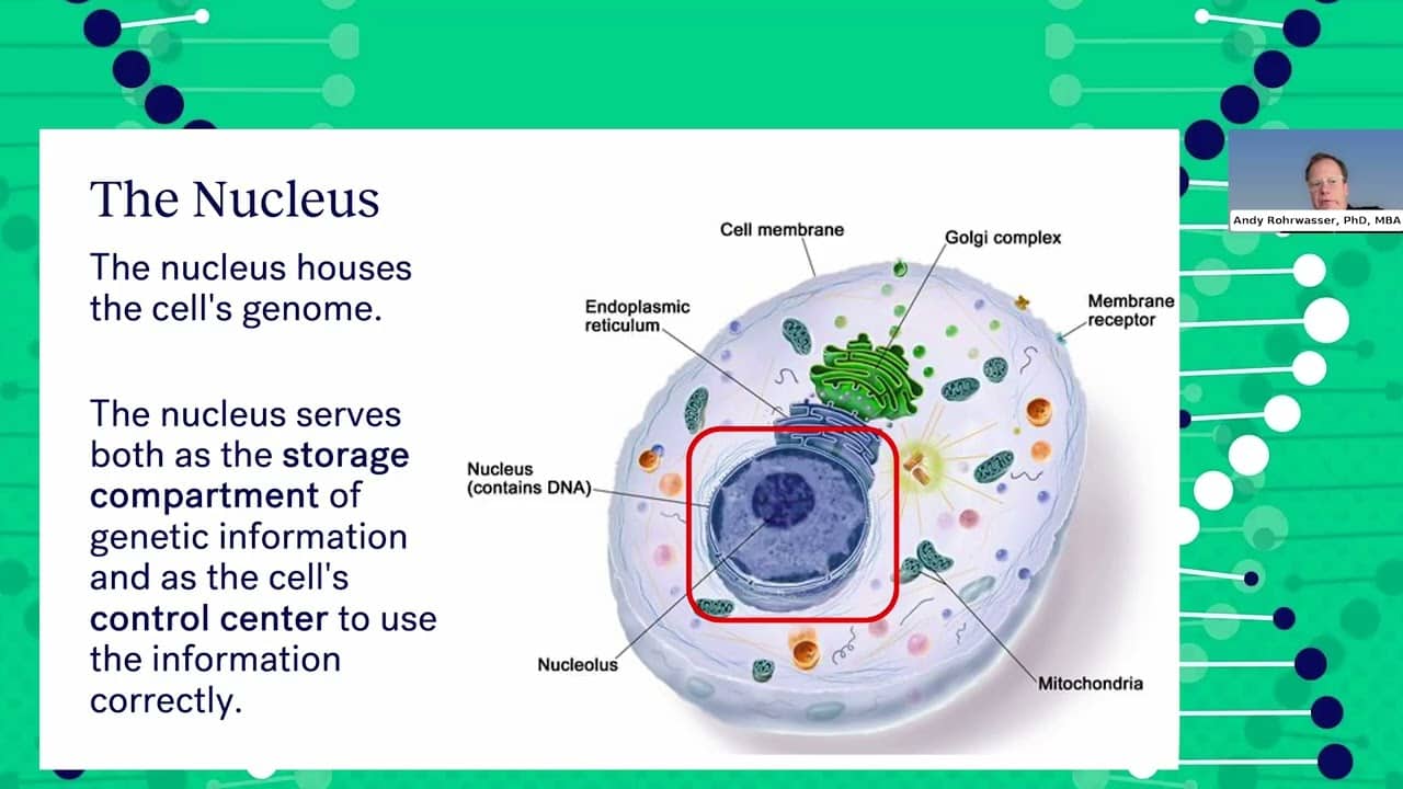
Myotonic dystrophy (DM) is a genetic condition caused by abnormal expansions of repeated DNA sequences that disrupt how cells function. DM is inherited in an autosomal dominant pattern, meaning a parent with the mutation has a 50% chance of passing it on to each child.
In DM type 1 (DM1), the condition is caused by an expansion of CTG repeats in the DMPK gene on chromosome 19. While most people have a small number of repeats, larger expansions lead to disease. These repeats can increase in size as they are passed from one generation to the next, a phenomenon known as genetic anticipation, often resulting in earlier onset and more severe symptoms in children. Very large expansions can cause congenital DM, which typically occurs through maternal transmission.
In DM type 2 (DM2), the condition is caused by an expansion of CCTG repeats in the CNBP gene on chromosome 3. DM2 generally does not show anticipation to the same extent as DM1, and symptoms usually begin in adulthood.
Because repeat size, inheritance patterns, and symptom severity can vary widely, genetic counseling plays a critical role in diagnosis, family planning, and helping individuals and families understand their risks and options.
Quick facts
Genetics – DM1
Patterns:
- DM1 is caused by the expansion of an unstable CTG repeat sequence in an untranslated, but transcribed, portion of the 3’ untranslated region of the dystrophia myotonica protein kinase (DMPK) gene located on chromosome 19q13.3.
- The normal number of CTG repeats in this region is 5 to 37. Repeat numbers greater than 50 are considered diagnostic of DM1. Occasionally, individuals are identified as inheriting 37 to 49 CTG repeats. Repeats of this length may be encountered in the side branches of known DM1 families, particularly in the older generations, or occasionally by chance in the general population. Individuals with 37 to 49 CTG repeats have not been reported to date to develop detectable DM1 symptoms. However, such “pre-mutations” can expand into the disease range in subsequent generations, particularly when transmitted by men.
- A diagnosis of DM1 in one person in a family has implications for other family members, giving rise to questions about whether or not the affected person should tell family members who show no symptoms and then whether or not those family members should be tested. Diagnosis of DM1 in a presymptomatic person (including a child) can have important implications for health monitoring and family planning, but it can also raise the possibility of difficulty in obtaining insurance or encountering prejudice in the workplace.
Symptoms:
- The diagnosis of DM1 should be suspected in anyone presenting with at least three of the following:
- Eyelid ptosis.
- Distal weakness, primarily of the finger and wrist flexors, without contractures.
- Myotonia or “stiffness” of muscles.
- Pre-senile cataracts, especially the polychromatic type.
- The diagnosis of DM1 should be suspected in anyone presenting with any one of the above or a family history and:
- First-degree heart block.
- Irritable bowel syndrome (IBS) or elevated liver enzymes.
- Gallstones at a young age.
- Prolonged recovery or respiratory arrest following an anesthetic.
- Insulin resistance or diabetes.
- Hypogonadotrophic hypogonadism.
- Excessive daytime sleepiness (EDS).
- Mild learning difficulty.
Diagnosis:
- Discuss the following tests with your doctor:
- While DNA testing, including prenatal and presymptomatic testing, for DM1 is now widely available, there are many potential pitfalls in interpreting the results for the patient and family, making genetic counseling a useful part of the diagnostic process.
- DM1 test via molecular genetic testing is the first line of investigation for anyone suspected of having DM1. More than 50 CTG repeats in the 3’ untranslated region of the DMPK gene on chromosome 19 are considered to have DM1. False-negative genetic testing results can occur, even in a family with an established DM1 diagnosis; expert referral is recommended.
- Consider a referral to genetic counseling services or a neurologist with expertise in DM1, even if you don’t desire to have children.
- For physical findings that are suspicious for a diagnosis of DM1 via physical examination with particular emphasis on neuromuscular, cardiovascular and respiratory assessments, obtain a three generation family history.
Treatment:
- Refer to:
- Genetic counseling for those who exhibit clinical signs indicative of DM1, for at-risk family members, in order to enable them to make an informed decision about whether to proceed to genetic testing. Such testing should be done through an accredited laboratory experienced in providing DM1 diagnoses. Individuals with 37 to 49 CTG repeats are deemed very unlikely to develop detectable DM1 symptoms. However, such “premutations” can expand into the disease range in subsequent generations, particularly when transmitted by men. Individuals thus identified should be offered genetic counseling to discuss their risk for transmitting DM1.
- Neuromuscular disease specialist, most likely a neurologist or clinical geneticist with a particular interest in inherited neuromuscular disease, who can facilitate a primary “wholesystem” evaluation, prioritizing additional symptom-specific referrals, and providing ongoing clinical management of the condition.
- Cardiologist if significant cardiac symptoms are detected. Anyone suspected of having a diagnosis of DM1 should be immediately advised of the risks of anesthesia and sedation and assessed for possible cardiac complications.
- Review pedigree annually. Genetic counseling should be repeated when new information or circumstances change the risks for family members.
- Discuss and convey the complexities of the inheritance patterns observed in this disease, particularly the risk of a minimally affected mother giving birth to a severely affected child, via genetic counseling.
- Male and female DM1-affected individuals may have difficulty conceiving and that the difficulty increases with age.
- Mutation carriers should inform their close relatives of the possibility that they may also have inherited the risks and repercussions of DM1, even if they or their children are currently asymptomatic.
- Preimplantation genetic diagnosis can allow selective implantation of unaffected embryos. Prenatal diagnosis by amniocentesis or chorionic villus sampling can allow for termination of an affected pregnancy. It can also prepare the obstetric team for the birth of a DM1- affected baby.
Genetics – DM2
Patterns:
- DM2 is caused by the expansion of an unstable CCTG repeat sequence in intron 1 of the CNBP gene in chromosome 3q21.3. The normal number of CCTG repeats in this region is less than 28. Repeat numbers greater than 75 can be considered diagnostic of DM2.
Symptoms:
- Initial symptoms may relate to grip myotonia. Alternatively, myotonia may be inconspicuous, and the initial symptoms may involve weakness of muscles around the hips or shoulders.
- Common symptoms are difficulty standing up from a low chair, rising from the ground or a squatting position, or climbing stairs. Reaching up or working with the arms overhead also may be difficult. People with DM2 often experience unusual fatigue with exercise.
- Muscle pain in the neck, back, shoulders, hip flexors, and upper legs may be a prominent symptom
- Through this inherited genetic anomaly, individuals living with myotonic dystrophy type 2 can experience varied and complex symptoms, including:
- Pain
- Skeletal muscle problems
- Muscle weakness and cramping
- Heart complications
- Breathing difficulties
- Digestive problems
- Excessive daytime sleepiness
- Early cataracts
- Hormonal imbalances
- Speech and swallowing difficulties
- Diabetes
- Immune system responses
- Impaired vision
- Cognitive difficulties
Diagnosis:
- Discuss the following tests with your doctor:
- While DNA testing for DM2 is now widely available, there are many potential pitfalls in interpreting the results without help, making genetic counseling a useful part of the diagnostic process. A diagnosis of DM2 in one person in a family has implications for other family members, giving rise to questions about whether or not the affected person should tell family members who show no symptoms and then whether or not those family members should be tested.
- Diagnosis of DM2 in a presymptomatic person can have important implications for health monitoring and family planning, but it can also raise the possibility of difficulty in obtaining certain types of insurance or encountering prejudice in the workplace.
Treatment:
- Consider a referral to genetic counseling services or a neurologist with expertise in DM2, even for those who do not intend to have children.
- Review pedigree annually. Genetic counseling should be repeated when new information or circumstances change the risks for family members.
- Help mutation carriers inform their close relatives of the possibility that they may also have inherited the risks and repercussions of DM2, even if they or their children are currently asymptomatic.
Genetics – CDM
Patterns:
- DM1 is caused by the expansion of an unstable CTG repeat sequence in an untranslated, but transcribed, portion of the 3’ region of the dystrophia myotonica protein kinase (DMPK) gene located on chromosome 19q13.3.
- Repeat size is often large (typically >1000 repeats) but the repeat size cannot absolutely in isolation be used to determine whether a child will have CDM or how severe his/her symptoms will be.
- Once a family has had a child with CDM, there is an increased risk that the next child with DM1 will have congenital form as well.
Symptoms:
- Congenital DM1 (CDM) is defined in a child who has one or more of the following features:
- Physical signs or symptoms attributable to DM1 at birth, or in the first month of life, including one or more of the following features: respiratory failure, feeding problems, weakness and hypotonia, clubfoot, polyhydramnios, and/or reduced fetal movement.
- Genetic confirmation of expanded CTG repeat size.
- Need for medical intervention or hospitalization in the first month of life for medical issues specific to myotonic dystrophy. Diagnosis may not necessarily be made in the neonatal period but could be made later in life if the above criteria were demonstrably present.
- Maternal transmission bias is nearly always maternal and does not appear to be related to the severity of the disease in the mother. The mutated gene is only very rarely inherited from the father in newborns with myotonic dystrophy.
Diagnosis:
- Discuss the following tests with your doctor:
- DM1 in the pediatric age range (that do not meet the congenital criteria) are herein referred to as childhood-onset DM1. The diagnosis of childhood-onset DM1 can be made at any age if features of DM1 were demonstrably present during the childhood years but were not medically identified or diagnosed.
- There are other classification systems in the literature that further subdivide by age of symptom onset, such as the following: mild and severe congenital (age 0-1 years), childhood (1-10 years), and juvenile (10-18 years).
- Genetic counseling if clinical signs indicative of DM1 are present, to enable that an informed decision is made about whether to proceed to genetic testing. Such testing should be done through an accredited laboratory experienced in providing DM1 diagnoses (see myotonic.org). Individuals with 37 to 49 CTG repeats are deemed very unlikely to develop detectable DM1 symptoms. However, such “premutations” can expand into the disease range in subsequent generations.
- While DNA testing, including prenatal and presymptomatic testing for DM1 is now available, there are many potential pitfalls in interpreting the results without help, making genetic counseling a useful part of the diagnostic process.
Treatment:
- In many cases, a child with DM1 will be the first person in the family diagnosed with DM1, due to genetic anticipation. A diagnosis of DM1 in one person in a family has implications for other family members, raising questions about whether other family members who show no symptoms should be informed of the diagnosis and whether those family members should be tested. Genetic counseling for affected families should convey information about:
- The inheritance pattern of disease (autosomal dominant inheritance).
- The wide variability in the scope and severity of DM1 symptoms, even within the same family.
- The possibility of changes in symptom scope and severity over time.
- The likelihood that the mutation will expand and the disease will become more severe as it is passed from generation to generation (anticipation) and as individuals age.
- The possibility of a minimally-affected mother giving birth to a severely affected child.
- Options for family planning.
- Help mutation carriers inform their close relatives of the possibility that they may also have inherited the risks and repercussions of DM1, even if they or their children are currently asymptomatic.
- Do not use CTG repeat numbers, if available, for genetic advice or prognostication; these need to be discussed with a genetic counselor.
- Parents who have a child with myotonic dystrophy have a 50% risk of having another child with DM1, and clinical experience suggests that they are likely to have congenital or childhood-onset in future births as well.
- Suggest that parents consider in vitro fertilization with pre-implantation diagnosis to prevent DM1 transmission, or other alternatives for expanding their family.
- If the family and physician are considering testing an asymptomatic child, consider that all parties take part in a counseling session before testing, and at the time of the disclosure of the result. The counseling should involve the child, parents, child’s physician, a genetic counselor, and if necessary, a psychologist. This may be cumbersome and deter casual testing; at least consider this approach for critical cases.
- Once the diagnosis is confirmed, consult an expert multi-disciplinary myotonic dystrophy team to coordinate care, prioritize symptom management and make appropriate additional referrals.
How does repeat length relate to the severity of myotonic dystrophy?
It is important to understand how CTG repeat length is associated with the severity of myotonic dystrophy type 1. CTG is the type of trinucleotide repeat expansion found on the DPMK gene inherited by individuals with DM1. For individuals with myotonic dystrophy type 2, the expanded CCTG repeat is found in the CNBP gene.
- Individuals with mild or late onset DM1 typically have a CTG length of 50-150, often with age of onset over 50 years old with symptoms such as mild cataracts and mild weakness.
- Individuals with typical or adult onset DM1 have a CTG length of 150-1000, typically with age of onset in the teenage years and older, and symptoms such as early cataracts, weakness and myotonia.
- Children with childhood onset DM have a CTG length of 600-1200, with age of onset between 1 and 10 years old, commonly exhibiting symptoms such as intellectual impairment and GI distress.
- Babies with congenital onset DM have a CTG length of 800 or more, with age of onset at birth and exhibiting symptoms such as floppiness, breathing and feeding problems.
For individuals with adult-onset myotonic dystrophy type 2, in general, repeat lengths less than 28 are considered normal, while repeats greater than 75 up to 1,000 are associated with clinical symptoms such as myalgic pains, myotonia, hip and neck flexor muscle weakness, cataracts and cardiac arrhythmias.
For more information on the significance of CTG repeats, watch Dr. Darren Monckton’s presentation Everything You Wanted to Know About CTG Repeats.
Related videos
Ask-the-Expert: Genetics & myotonic dystrophy
Published: October 9, 2025
Understanding DM1: From Genetics to Care - 2025 MDF Conference
Published: August 28, 2025

DM2 Essentials: Understanding, Managing, and Thriving with DM2 - 2025 MDF Confer...
Published: August 28, 2025

Genetics Unveiled: Understanding Testing, Counseling & Family Conversation a...
Published: August 28, 2025